GentleWave

The Science Behind the GentleWave® Procedure

GentleWave Procedure IllustrationPatient-Centered Root Canal Treatment Technology

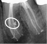
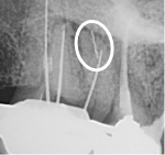
We developed the GentleWave® Technology in pursuit of improving the efficacy and efficiency of root canal treatment (RCT), as well as the patient experience. For well over a century, RCT has depended primarily on files to manually remove infected tissue and tooth structure from inside the tooth. Files, however, have limited reach inside complex root canal anatomy4 and can leave behind infected tissue3 and bacteria, leaving the tooth susceptible to reinfection.

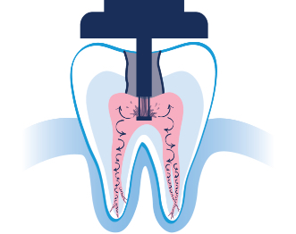
The minimally invasive1 approach of the GentleWave Procedure minimizes manual instrumentation1,3 and instead utilizes a powerful vortex of procedure fluids3 and acoustic energy that cleans the deepest, most complex portions of the root canal system.1,3

Redefining Standard Root Canal Therapy

During the GentleWave Procedure, fluids undergo a multistage optimization process that energizes and propels fluids throughout the root canal system1,3 to remove infected tissue, debris and bacteria.3 The result is a more efficient and effective clean that promotes early healing.2

GentleWave® Technology at Work

This short video demonstrates how optimized procedure fluids successfully clear canals of infected tissue (represented by the blue material inside the root canals).

Discover the Innovative GentleWave® Procedure

What makes the GentleWave Procedure the differentiator for patients who don’t want RCT? Here’s just a few reasons patients opt for today’s root canal alternative.

The Possible Complications in Manual Instrumentation

While files have been used successfully in RCT for many years, they have the potential to:

  • Remove too much tooth structure, which may weaken the remaining tooth2
  • Leave behind infected tissue3 due to limited reach in complex root canal anatomy4
  • Break off inside the tooth, where they often cannot be removed5
  • Puncture through the tooth, leaving the tooth unsalvageable6
  • Create "smear layer"7—a paste-like substance containing debris and bacteria
Complex Anatomy

Figure 1: Complex Anatomy. This lateral canal would be virtually impossible to clean using only files.

Separated Instrument

Figure 2: Separated Instrument. The circled area of this patient’s tooth shows a piece of a file left behind.

4 AEGIS Communications, By James Bahcall, DMD, MS. (n.d.). Today’s Endodontic Therapy Driven By Advances in Technology, Changes in Thinking. Retrieved from https://www.aegisdentalnetwork.com/cced/2015/05/todays-endodontic-therapy-driven-by-advances-in-technology-changes-in-thinking

5 Evaluation of the Factors and Treatment Options of Separated Endodontic Files Among Dentists and Undergraduate Students in Riyadh Area. (n.d.). Retrieved from https://www.ncbi.nlm.nih.gov/pmc/articles/PMC4843379/

6 Root perforations: Aetiology, management strategies and outcomes. The Hole Truth. (1985, May 1). Retrieved from https://www.researchgate.net/publication/297736502_Root_perforations_Aetiology_management_strategies_and_outcomes_The_hole_truth

7 Violich DR and Chandler NP. (n.d.). The smear layer in endodontics - a review. - PubMed - NCBI. Retrieved from https://www.ncbi.nlm.nih.gov/pubmed/20002799